Your eye doctor stresses the importance of having vision tests done but your question is why?

Glaucoma is a disease that damages your optic nerve. Primary open angle glaucoma is when the eye does not drain fluid like it should. As a result, eye pressure builds up causing high pressure and damages the optic nerve. Open angle glaucoma is painless and causes no vision changes at first. Most people don’t notice small changes but once they do notice a decrease in vision due to glaucoma, the damage is done, so getting tests done that shows gradual vision changes is very important. When managing glaucoma there are many reasons ophthalmologists/optometrists order tests. One reason is to establish a baseline so that later it is possible to tell if something has changed. Some vision tests are done to determine a specific problem or diagnosis.
The visual field test is a subjective measure of central and peripheral vision and is used by your doctor to diagnose, determine the severity of, and monitor your glaucoma. It evaluates vision loss, damage to the visual pathways of the brain, and other optic nerve diseases. The most common visual field test uses a specific light stimulus to repeatedly move around the patient’s peripheral vision. During the test the patient puts their head positioned in the machine and clicks a button each time they see a light. Each eye is tested separately. Imaging of the optic nerve is complementary to visual field testing and using both together is useful. These images are important because they give detailed documentation of the appearance of the optic nerve.

Your doctor may recommend retinal testing if you have a certain disease or condition such as diabetes or macular degeneration. Diabetes can damage the blood vessels in your retina and if not controlled can cause vision loss. AMD (age related macular degeneration) is a condition that causes loss in the center of vision. Nonexudative macular degeneration also known as dry AMD usually starts with the appearance of yellow-colored spots in the macula caused by the build-up of fatty deposits called drusen. Exudative macular degeneration also known as wet macular degeneration is when abnormal blood vessels grow under the retina. These vessels may leak fluids causing scarring of the macula. Dry AMD can lead to wet AMD which may require treatment. Many people don’t realize they have AMD until their vision is very blurry. Your ophthalmologist can look for early signs of this before you notice change in your vision.
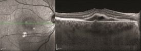
There are several different types of retinal tests such as fundus photos, Optical coherence tomography (OCT), Fluorescein angiography (FA) and electroretinography (ERG). Photos of the back of the eye are used to diagnose and document progression and treatment of eye conditions such as macular degeneration, diabetic retinopathy, and other conditions involving the retina. OCT is used to capture images of the retina to help diagnose epiretinal membranes, macular holes, macular swelling, to monitor age-related macular degeneration, and to monitor response to treatment. The FA is a test that uses a dye to make the blood vessels in the retina stand out under a specific light. This helps to identify closed blood vessels, leaking blood vessels, abnormal blood vessels, and changes to the back of the eye. An ERG test measures the electrical response of the light-sensitive cells in your eyes. These cells are rods and cones and form the back of the eye called the retina. All these tests are painless and give your doctor a better idea of the health of your eyes. It is very important to keep up with your appointments and to keep on a regular schedule with your eye doctor.
Bond Eye Associates is accepting new patients in both of their locations: Peoria and Pekin, IL. Please call Bond Eye Associates to schedule your yearly health vision exam with confidence knowing that they have been a trusted, locally owned, medical practice for over 40 years at 309-692-2020 or click here.


